No special preparation is needed. You can eat, drink, and take your regular medications normally prior to the test.
Non-Invasive CARDIOLOGY
2D & 3D Echocardiography
Ultrasound imaging to understand how your heart pumps and its valves work.
Book a consultation About this treatment ↓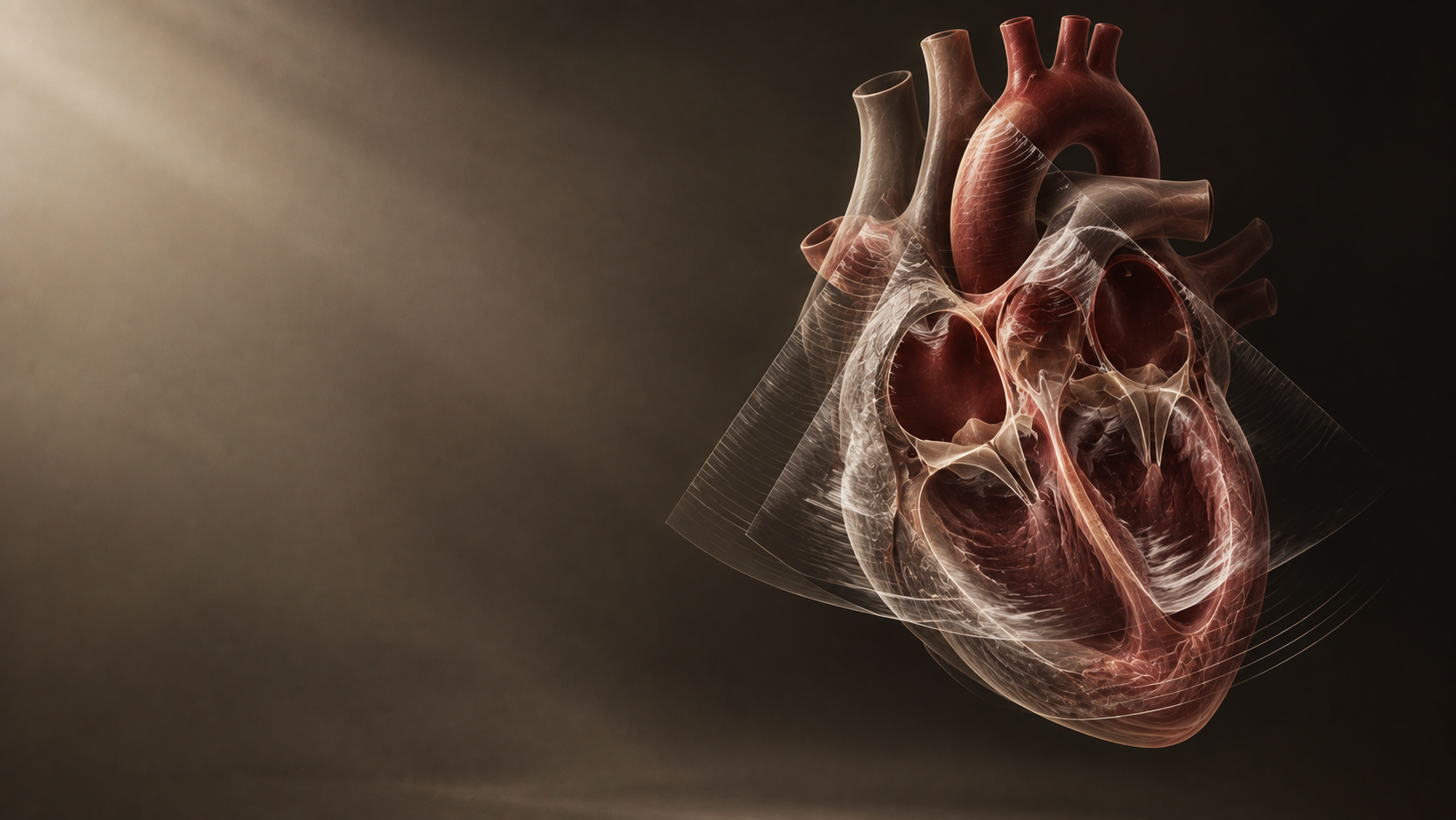
- The focus
- Heart function & valves
- The approach
- Non-invasive ultrasound
- Your next step
- Personal assessment
ABOUT THIS TREATMENT
What you need to know.
Non-invasive cardiac ultrasound evaluating ejection fraction, valve motion, heart wall movement, and congenital structural anomalies.
01Understanding the treatment
Echocardiography uses sound waves to generate live high-definition moving pictures of the heart. Dr. Sandhu utilizes 2D/3D Color Doppler Echocardiography to evaluate heart muscle contractility, pump strength (Ejection Fraction), valve narrowings or leakages, and pericardial fluid.
02Who may benefit?
- Patients with unexplained breathlessness, swollen ankles, or fatigue
- Individuals with heart murmurs detected during physical examination
- Patients with long-standing hypertension or suspected valve disease
- Routine follow-up for patients post-angioplasty or valve surgery
03What happens during treatment?
Pain-free 20-minute scan performed by applying an ultrasound transducer over the chest wall.
04Recovery & follow-up
No downtime. Instant diagnostic report generation.
05Benefits of this approach
- 100% non-invasive with zero radiation exposure
- Accurate measurement of cardiac pumping fraction (EF %)
- Clear visualization of all four heart valves in real time
A LITTLE MORE CLARITYYour questions,